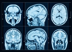
Parkinson’s disease (PD) is typically diagnosed based on clinical exam, but there are imaging tests that are available to help in diagnosis. The most commonly known in the US is the Dopamine Transporter Scan (DaTscan), an imaging test in which a radioactive tracer, Ioflupane (123I) — which is also called DaTscan — is injected into the blood, where it circulates around the body and makes its way into the brain. It attaches itself to the dopamine transporter, a molecule found on dopamine neurons. Several hours after the tracer has been injected, special imaging equipment scans the head to detect the presence of DaTscan. A DaTscan in someone with PD typically shows a smaller signal in a part of the brain called the striatum, where the ends of the dopamine neurons are meant to be. Learn more about DaTscan for Parkinson’s
Beyond DaTscan – other Parkinson’s disease brain imaging tests
There are a number of other imaging tests that can help shed light on whether a person does or does not have PD. Each of these tests have pros and cons which we will discuss below. Some of these tests can currently be performed clinically, but some are research tools only. In either case, most of these tests are not currently approved specifically for the diagnosis of PD, so they are not covered by insurance.
As more data is gathered about these tests and their utility in PD, the hope is that they will become more widely accessible. If you are interested in exploring any of the currently available testing options, please speak with your neurologist.
FDG-PET (Fluorodeoxyglucose Positron Emission Tomography)
How it Works: FDG-PET measures how much glucose is used by different parts of the brain. Since the brain cells need glucose to function, this scan shows patterns of brain activity. PD often causes reduced glucose metabolism in parts of the brain like the parietal and occipital lobes, while the motor regions are relatively preserved. These changes are distinct from those seen in similar conditions like multiple system atrophy (MSA) or progressive supranuclear palsy (PSP).
Evidence: NeuroImage: Clinical outlined a study that validated a specific PD-related metabolic brain pattern (PDRP) using FDG-PET scans. The study found that the scans were able to reliably distinguish early-stage PD patients from healthy controls correlated with clinical measures supporting its potential as an objective biomarker for tracking disease progression and treatment response.
Strengths: Well-established, high accuracy, ability to distinguish between different parkinsonian syndromes
Limitations: Does not directly measure dopamine, involves exposure to radiation, and can be expensive
F-DOPA PET (Fluorodopa Positron Emission Tomography)
How it Works: F-DOPA PET uses a radiolabeled version of the amino acid L-DOPA (the precursor to dopamine) to image dopamine-producing cells within the brain. PD patients typically show reduced uptake of F-DOPA in the striatum. This usually occurs asymmetrically on the side opposite of the first displayed symptoms. This reduction reflects the loss of dopaminergic neurons that can be differentially detected by this imaging.
Evidence: A study published in 2016 in The American Journal of Nuclear Medicine and Molecular Imaging found that in 27 movement disorder patients, qualitative and quantitative F-DOPA PET scans accurately distinguished PD from other motor disorders, achieving a sensitivity of 95.4% and specificity of 100% with no false positives and one false negative.
Strengths: High resolution and sensitivity, helpful for early-stage disease detection, approved by the FDA for use in diagnosing parkinsonian syndromes
Limitations: Limited availability, expensive, and requires specialized facilities
Transcranial Sonography (TCS)
How it Works: TCS utilizes ultrasound waves through the skull to visualize deep brain structures like the substantia nigra. Many PD patients show a brighter (hyperechogenic) area in the substantia nigra compared to healthy individuals.
Evidence: A large TCS study published in 2023 analyzed the TCS scans of 854 PD patients and 775 healthy controls and concluded this diagnostic tool could distinguish PD from healthy controls with 88% accuracy, performing better than that of traditional diagnostic methods.
Strengths: Inexpensive, easily accessible, portable, quick, and no radiation exposure
Limitations: Lack of specificity, not possible with all patients, difficult standardization for analysis of data
Alpha-Synuclein Imaging
How it Works: The pathologic hallmark of PD is the accumulation of a misfolded protein called alpha-synuclein in the brain cells. A variety of new imaging agents are being developed to detect this protein directly in the brain and circulating in the blood.
Evidence: A 2024 CellPress publication illustrated a novel small molecule ligand (C05-05) for imaging of alpha synuclein aggregates using PET in mice. While this work is still preclinical, models like this are essential to prove translatable diagnostic methods for early PD diagnosis and monitoring.
Strengths: Potential for early, definitive diagnosis
Limitations: Still under research where specificity, safety, and practical use remain hurdles for useful and accurate detection
Can an MRI detect Parkinson’s disease?
Standard MRI scans cannot directly detect PD. However, specialized MRI techniques are showing promise as diagnostic tools and are even used routinely in some parts of the world.
Nigrosome-1 imaging on SWI (Susceptibility Weighted Imaging) MRI
How it Works: The substantia nigra pars compacta is the part of the brain that contains a high density of dopaminergic neurons that degenerate in PD. It can be further subdivided into a matrix (60%) and five clusters of dopaminergic neurons called nigrosomes (40%). The dopaminergic neurones in nigrosome-1 are the first to degenerate in PD. Nigrosome-1 can be visualised on magnetic resonance imaging (MRI) at 3 Tesla using a sequence called susceptibility weighted imaging (SWI). Nigrosome-1 is normally a bright structure located between two dark linear regions. This normal appearance has been likened to the shape of a swallow’s tail and has been called the “swallow tail sign”. In PD, the bright nigrosome-1 is lost and thus there is the absence of the “swallow tail sign”. Nigrosome-1 SWI MRI is used routinely as part of the diagnostic workup of PD in some parts of the world, for example, Australia.
Evidence: A systematic review from 2020 collected data from nineteen studies covering over 1500 subjects. The overall sensitivity was 0.94, and the overall specificity was 0.90.
NM-MRI (Neuromelanin-Sensitive Magnetic Resonance Imaging)
How it Works: Neuromelanin is a darker pigment found in healthy dopamine neurons in the substantia nigra (the region of the brain affected by PD). NM-MRI uses special MRI settings to detect this pigment. PD typically shows reduced signal intensity and volume in the substantia nigra which is consistent with neuron loss and PD symptoms.
Evidence: A study published in Nature in 2024 analyzed NM-MRI scans within the substantia nigra of 21 PD patients along with 13 essential tremor (ET) patients and 18 healthy controls. The authors concluded that the NM-MRI scans could distinguish PD patients from both ET and healthy groups with 100% sensitivity and 96-99% specificity. The study also found that the NM-MRI could even differentiate PD subtypes based on region-specific neuromelanin loss.
Strengths: Non-invasive, no radiation exposure, and MRI is widely accessible
Limitations: Still in early stages and experimental, image quality and interpretation vary between centers and clinicians.
Iron-Sensitive MRI Quantitative Susceptibility Mapping (QSM)
How it Works: QSM detects tiny magnetic changes in the brain caused by iron deposits, which accumulate in the substantia nigra in PD. QSM within PD patients typically shows increased iron in the substantia nigra, which may correlate with disease severity and progression.
Evidence: A review published in NeuroImage 2024 gathered the data in support of using Iron-Sensitive MRI QSM to detect elevated iron levels in PD patients as an aid in diagnosis.
Strengths: Non-invasive, no radiation exposure, and MRI is widely available.
Limitations: Currently only used in research and the interpretation of the data can be complex and difficult to standardize for diagnosis
Other Advanced MRI Techniques
Diffusion Tensor Imaging (DTI)
This imaging maps white matter pathways in the brain. PD is associated with disrupted connections (near substantia nigra) and this technique could be useful for early detection when used in combination with other imaging.
Resting-State fMRI
This type of MRI looks at how different brain regions communicate when the patient is at rest and not performing a specific task. PD patients show altered brain network connectivity which may relate to symptoms like cognitive changes and depression.
Magnetoencephalography (MEG)
This captures brain wave activity in real time but is in the very early experimental stages for PD. MEG may be able to reveal abnormal rhythms in motor areas and could help monitor treatment effects.
Combining Imaging Methods
Each one of these technologies offers a unique way of analyzing the brain to help diagnose PD whether it focuses on structure, function, dopamine levels, or protein buildup. Using two or more of these technologies together can provide a more complete picture for accurate diagnosis and progression of the disease. Here are some combinations that could prove vital for enhanced diagnosis and treatment of PD:
- F-DOPA + MRI: Help pinpoint where dopamine loss is greatest within the structure of the brain as defined by MRI
- FDG-PET + DTI: Could distinguish PD from other disorders by combining metabolic and structural data
- NM-MRI + QSM: Could show both neuron loss and iron buildup
- Alpha-Synuclein PET + FDG-PET Imaging: May provide the best sensitivity and precision for early detection and treatment
It is important to note that while these new and emerging techniques seem promising, specific combinations and certain technologies are still in their early stages of development and will need to be evaluated in clinical trial before becoming widely available for diagnosis in PD.
Tips & Takeaways
- Imaging tools beyond DaTscan are being developed to help in the diagnosis of PD and related disorders
- FDG-PET and F-DOPA-PET are powerful tools, with proven accuracy in early diagnosis and disease differentiation
- Advanced MRI methods like nigrosome-1 SWI imaging, NM, and QSM are promising, non-invasive options for identifying brain changes without radiation
- Transcranial ultrasound is a low-cost option that may help in early screening or in places without access to high-tech scanners
- Alpha-synuclein imaging is on the horizon and could transform how we diagnose and treat PD
- Combining different imaging tools offers the best results, helping doctors build a more complete picture of disease activity and progression
This blog was written by Clark Jones, PhD, and was reviewed, edited, and approved by Dr. Rebecca Gilbert.